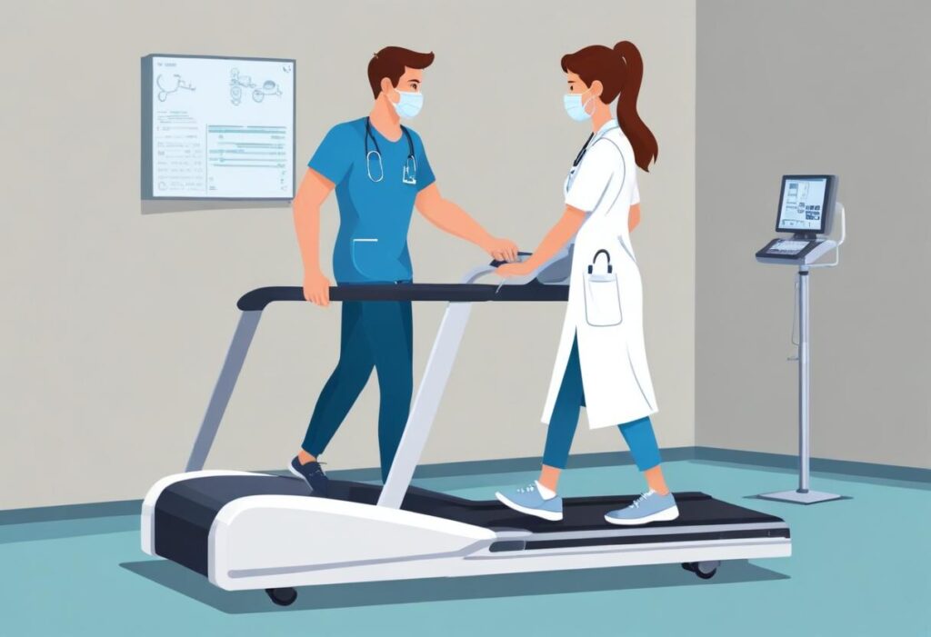
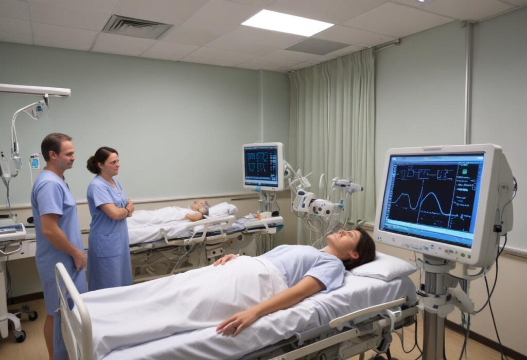
Initial tests may include an electrocardiogram (ECG) while resting, and again while working or running on the treadmill or a cycle ergometer. If these tests are positive for reduced blood supply to your heart muscle, then the person may have to undergo “cardiac catheterization or Coronary Angiography. The risk of angiography in well-established centers is less than 0.01%.
What is ECG (Electrocardiogram):

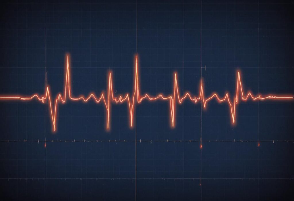
ECG is a graphical recording of the electrical activity of the heart. The abnormal electrical wave pattern of the heart can be picked up and heart disease can be diagnosed. Resting ECG is no more a gold standard for diagnosing Acute Heart Attack because in some diseases ECG may not accurately diagnose the heart disease for example Diabetic people may not show changes in ECG (termed as silent heart attacks). It’s a non-invasive procedure that involves placing electrodes, usually in specific locations on the chest, arms, and legs. These electrodes detect the electrical signals generated by the heart as it contracts and relaxes. During an ECG, the electrodes pick up these electrical impulses and transmit them to the ECG machine, which displays the heart’s activity as a series of waves on a graph or monitor. Each wave represents a different phase of the heart’s electrical cycle: The P wave represents atrial depolarization (contraction of the atria). The QRS complex represents ventricular depolarization (contraction of the ventricles). The T wave represents ventricular repolarization (relaxation of the ventricles).
The ECG provides valuable information about the heart’s rhythm, rate, and overall electrical activity. It helps diagnose various heart conditions, such as arrhythmias (abnormal heart rhythms), heart attacks, conduction abnormalities, and other cardiac disorders.
ECG is a less expensive test commonly used in routine check-ups, during medical emergencies, before surgeries, or when heart-related symptoms are present, such as chest pain, palpitations, shortness of breath, or dizziness. They’re quick, painless, and provide essential insights into heart health, aiding healthcare providers in making accurate diagnoses and determining appropriate treatments or interventions.
Treadmill Test (stress test):

This test helps to identify effort tolerance, stratify the risk of heart attack, and cardiac arrhythmia, etc. A Treadmill Test, commonly known as a stress test or exercise stress test, is a diagnostic procedure used to evaluate the heart’s response to physical activity, typically performed on a treadmill or a stationary exercise bike.
During a treadmill test
Preparation:Electrodes are placed on the chest to monitor the heart’s electrical activity (ECG or EKG). Baseline measurements of blood pressure, heart rate, and ECG are recorded while the patient is at rest.
Exercise: The patient starts walking on the treadmill or pedalling the stationary bike. The exercise intensity gradually increases, either by increasing the speed, incline, or resistance.
Monitoring: Throughout the test, the patient’s heart rate, blood pressure, and ECG are continuously monitored to assess how the heart responds to exertion.
Symptom Assessment: The patient is encouraged to exercise until they reach a target heart rate, experience symptoms such as chest pain or fatigue, or until the healthcare provider decides to stop the test due to certain ECG changes.
The goal of a stress test is to provoke and evaluate the heart’s response to increased physical activity, mimicking the stress the heart experiences during exercise. It helps in:
Detecting abnormal heart rhythms (arrhythmias) that may occur during exertion.
Assessing blood flow to the heart.
Evaluating exercise tolerance and identifying symptoms such as chest pain or shortness of breath.
Determining the presence of coronary artery disease or other heart-related conditions:
The test is stopped if the patient experiences significant symptoms, abnormal changes in blood pressure or ECG, or reaches the maximum exercise capacity determined by the healthcare provider. Stress tests are useful in diagnosing heart conditions, assessing cardiovascular fitness, and guiding treatment decisions.
Echocardiogram (2D Echo)
It is an imaging technique of the heart by ultrasound. With this, one can study the structure and function of the heart muscles, valves, and anatomical details of the heart. However, presence of the coronary artery disease can not be diagnosed by this test and the effect of coronary artery disease on the heart muscle can be ascertained. An echocardiogram is a non-invasive and painless imaging test that uses high-frequency sound waves (ultrasound) to create detailed images of the heart’s structure, valves, chambers, and blood flow patterns.

During an echocardiogram
Transducer Placement:A technician places a small handheld device called a transducer on the chest. The transducer emits sound waves and captures their echoes as they bounce off the heart’s structures.
Image Generation: The sound waves produce real-time moving images of the heart on a monitor. These images display the heart’s walls, chambers, valves, and blood flow.
Doppler Imaging: Doppler ultrasound, often part of the echocardiogram, analyzes the speed and direction of blood flow within the heart, providing information about valve function and blood flow patterns.
Types of echocardiograms include:
Transthoracic Echocardiogram (TTE): This is the most common type, performed by placing the transducer on the chest wall to capture images of the heart’s structures.
Transesophageal Echocardiogram (TEE): In this procedure, the transducer is passed into the esophagus to obtain clearer and more detailed images of the heart, especially for evaluating certain heart conditions or when a TTE is insufficient.
Echocardiograms help in:
Assessing heart chamber size, thickness, and overall function.
Evaluating heart valve structure and function.
Detecting abnormalities in the heart’s structure or movement, such as congenital heart defects or heart muscle abnormalities. Diagnosing heart conditions like heart failure, heart valve diseases, pericardial diseases, and more. Echocardiography is a valuable tool for cardiologists and healthcare providers to evaluate and diagnose various heart-related conditions, providing detailed and real-time images that aid in treatment decisions and monitoring cardiac health
Nuclear Cardiology
With the help of radioactive agents, the structure and function of the heart disease can be assessed. Areas of the heart muscle at risk of ischaemic injury can be evaluated and the ventricular function can be known. Nuclear cardiology imaging uses small amounts of radioactive substances, known as radiotracers or radiopharmaceuticals, along with imaging techniques to assess the structure and function of the heart, these radiotracers are introduced into the body, often through injection, and they emit gamma rays that can be detected by specialized cameras called gamma cameras or SPECT (single-photon emission computed tomography) cameras. These cameras create detailed images of the heart, providing information about blood flow, heart muscle function, and overall cardiac performance.
Common nuclear cardiology procedures include:
Myocardial Perfusion Imaging (MPI): This test evaluates blood flow to the heart muscle. The patient receives a radiotracer that travels through the bloodstream and is taken up by the heart muscle. The gamma camera captures images of the heart at rest and during stress (such as exercise or medication-induced stress), allowing comparison to identify areas with reduced blood flow.
Cardiac PET (Positron Emission Tomography): PET imaging uses different radiotracers than SPECT scans and provides more detailed metabolic information about the heart. It can assess blood flow, oxygen use, and metabolism in heart tissue.
Gated Cardiac Blood Pool Imaging:This technique assesses the heart’s pumping function by tracking the movement of blood through the heart chambers. It can evaluate the heart’s chambers, valves, and overall function. Despite involving the use of radioactive materials, the doses used are considered safe and have minimal risks when performed under proper medical supervision.
Angiogram (Cardiac catheterization):
This is the gold standard test to know the anatomy of the coronary arteries and chambers of the heart. By using a catheter to inject x-ray contrast dye into the coronary arteries, good pictures of coronary artery tree can be obtained. With this one can make out the degree of the block, the location of the block, heart muscle and valve functions, and the severity of the disease. This information is vital for deciding the best possible treatment plan.
CT angiography (CTA) is a diagnostic imaging technique that uses computed tomography (CT) scanning to visualize and evaluate blood vessels throughout the body, particularly focusing on arteries. It provides detailed, cross-sectional images that offer valuable information about the structure and function of the arteries.
During a CT angiography procedure, a contrast material (often iodine-based) is injected into a vein to enhance the visibility of blood vessels. As the contrast material travels through the bloodstream, the CT scanner rapidly takes images, creating detailed pictures of the arteries. These images help in identifying any narrowing, blockages, or abnormalities in the blood vessels, providing information about blood flow and detecting conditions such as atherosclerosis, aneurysms, or blood vessel malformations.
CT angiography is commonly used to assess various parts of the body, including the brain, heart, lungs, abdomen, and extremities. In cardiac CT angiography, for instance, it is utilized to examine the coronary arteries and evaluate the presence of coronary artery disease or blockages.
This non-invasive imaging technique is valuable in diagnosing vascular conditions, guiding treatment decisions, and monitoring the effectiveness of treatments such as stent placements or bypass surgeries. Additionally, it allows for precise planning of interventions when needed.
Holter Monitoring
With this system, one can record the heart rate throughout the day, and helpful in special situations. Holter monitoring is a diagnostic test that continuously records the heart’s electrical activity over an extended period, typically 24 to 48 hours or even longer. It’s named after Dr. Norman J. Holter, who pioneered portable electrocardiogram (ECG) devices.
During a Holter monitoring test, small electrodes are attached to the chest, connected by wires to a portable recording device worn on a belt or shoulder strap. This device continuously records the heart’s electrical signals (ECG) as the person carries out their daily activities.
The purpose of Holter monitoring is to capture and assess heart rhythms and electrical activity over an extended period, allowing doctors to evaluate the heart’s behavior during normal activities, including sleep, exercise, stress, or periods of symptoms like palpitations, dizziness, or fainting. This continuous monitoring helps detect irregular heartbeats (arrhythmias) that may not be captured during a brief ECG in a doctor’s office.
After the monitoring period, the device is returned to the healthcare provider’s office, and the recorded data is analyzed to identify any irregularities or abnormalities in the heart’s rhythm. This information aids in diagnosing various heart conditions, such as atrial fibrillation, bradycardia, tachycardia, or other arrhythmias, guiding appropriate treatment decisions or further diagnostic tests.
